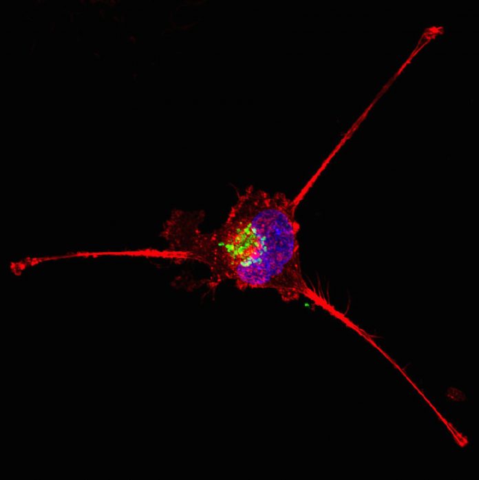
A recent study by researchers at the Baylor College of Medicine has shed light on the role of DAPK3 in triple-negative breast cancer (TNBC), detailing its regulation of epithelial-mesenchymal transition (EMT) which is a process linked to cancer metastasis. This research, published in the journal PNAS Nexus, provides a better understanding of the aggressive nature and the difficulty of treating TNBC, a subtype of breast cancer characterized by a lack of three common receptors: estrogen, progesterone, and the HER2/neu gene.
According to the American Cancer Society, TNBC accounts for about 15% of all breast cancer cases, with metastasis occurring in approximately 30–50% of these patients within five years of diagnosis. Given the aggressive behavior of TNBC, understanding the mechanisms driving its metastasis is crucial.
Kinases, whose expression is often altered in cancer, have been successfully targeted for treating other forms of cancer, with several kinase inhibitors already approved for use by the U.S. Food and Drug Administration (FDA). The Baylor team sought to discover what, if any, value these approved drugs might have in the treatment of TNBC.
“The challenge was to identify the kinase among hundreds of kinases in TNBC cells that could potentially give us an upper hand against this cancer,” said Junkai Wang, PhD, a former member of the lab of Charles Foulds, PhD, at Baylor which has been focused on better understanding TNBC and its vulnerabilities that could lead to better treatments.
To do this, the team used a lab test developed in-house called the kinase inhibitor pull-down assay (KIPA), a rapid method of kinase identification of hundreds of potential therapeutic candidates.
The team used 16 patient-derived xenografts (PDXs) to grow human breast cancer in immunocompromised mouse models. They then used KIPA to search for kinases whose quantity was significantly altered in cancer cells compared with normal cells.
“We found that TNBC cells have more of the kinase called Death-Associated Protein Kinase 3 (DAPK3),” Wang said. “We confirmed this finding in TNBC cell lines and in tumors.”
An important finding of this research was that while the protein levels of DAPK3 were high in TNBC, the levels of its precursor mRNA were not. The mRNA molecule carries the genetic sequence of the DAPK3 gene which is used by the cell to produce the protein. The significance of their finding is that many studies have relied upon mRNA data to figure out which proteins the cells produce. “If we had just looked into mRNA and not the protein levels in TNBC, we would have missed that the DAPK3 protein is overproduced in this cancer and deserves further attention,” Wang noted.
Additional work demonstrated that knocking out the DAPK2 gene to eliminate the DAPK3 protein did not affect cell growth, but it did prevent the migration and invasion of TNBC cells in the lab. When DAPK3 was knocked out in TNBC cells grown as tumors in the immunocompromised mice, the researchers observed no significant effect on tumor metastases, though the team noted that additional research is needed to confirm these findings.
The team also found how DAPK3 mediates its migration. “We found that DAPK3 reduces the levels of desmoplakin (DSP), a protein that is involved in regulating cell adhesion, which is related to a cell’s ability to migrate,” Wang said. “In addition, we discovered that a protein called LUZP1 binds to DAPK3, which protects it from being destroyed by the cell.”
Added Foulds: “Altogether, our findings have improved our understanding of what kinases control the spread of TNBC cells. We previously thought that a driver of cancer may affect cell proliferation and migration. But we found that in TNBC, DAPK3 does not regulate growth, but controls migration and invasion.”
The next steps for the Foulds lab will be additional research to learn more about how the DAPK3/LUZP1 complex functions to promote TNBC cells to migrate and to discover whether it is a viable therapeutic target.


