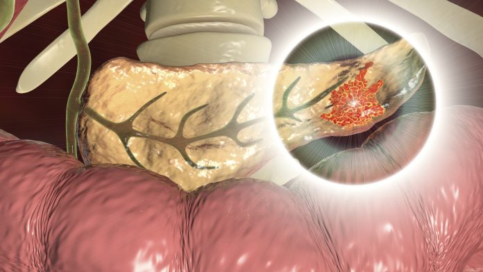
A new study led by researchers from the Champalimaud Foundation in Lisbon, Portugal, has, for the first time, detected pre-cancerous lesions in the pancreas using diffusion tensor imagining (DFI). Reporting on their work in the journal Investigative Radiology, the investigators detailed how they used DFI, a specialized form of MRI to detect pancreatic intraepithelial neoplasia (PanIN), precursor lesions to the development of pancreatic ductal adenocarcinomas (PDACs).
Unlike some cancers, the symptoms of pancreatic cancer that include stomach pain, weight loss, new-onset diabetes, and others are often confused with the onset of other health conditions. Further, pancreatic cancer lacks reliable non-invasive measures for diagnosis. This new capability using DFI is critical for both detecting these pre-malignant lesions and for better understanding PanIN biology and its role on PDAC.
“PanINs are not diagnosed by current imaging modalities. There is an urgent need for developing imaging methods for PanIN diagnosis and characterization,” the researchers wrote.
PDAC originates from precursor lesions known as PanINs in most cases. These lesions typically develop years before malignant PDAC manifests. The inability to detect PanINs has limited research into how pancreatic cancer develops, as well as efforts to develop treatments targeting the disease in its early stages.
To address this, the team turned to DTI, which has been used to study brain tissue for the past 30 years. The researchers in the Shamesh lab adapted DTI to detect the microstructural changes associated with PanINs in the pancreas.
“It’s not a new method—it was just never applied in the context of pancreatic cancer precursor lesions”, said Noam Shamesh, PhD, lead investigator and head of the preclinical MRI lab at Champalimaud Research. “It was Carlos [Bilreiro, MD, first author of the study] who came to see me with this idea.” Bilreiro is a doctor at the Champalimaud Clinical Centre’s Radiology Department.
DTI works by measuring the diffusion of water molecules within tissues. The movement of these molecules can reveal detailed information about the tissue’s microstructure, which in the case of PanINs, changes as the lesions develop. In their study, the researchers used DTI to capture high-resolution images of pancreatic tissue from transgenic mice genetically predisposed to developing PanINs. They then compared these images with histological samples, which showed that DTI could reliably detect the presence of these pre-cancerous lesions.
Building on these results, the team then tested their method on human pancreatic tissue samples. “We obtained human samples and demonstrated that DTI was just as effective in detecting lesions in human tissue as it was in mice,” Shemesh said.
While these results are promising, further studies are needed before DTI can be used in clinical practice. The main hurdle to clinical adoption is that the resolution of MRI scanners typically found in the clinic provide lower resolution images.
Building in this finding, the research team will conduct clinical trials to test DTI in human patients. The researchers are optimistic about the potential to refine the technique for clinical use, possibly combining DTI with other diagnostic methods like liquid biopsies or artificial intelligence to improve the accuracy and specificity of PanIN detection.


![Best Weight Loss Supplements [2022-23] New Reports!](https://technologytangle.com/wp-content/uploads/2022/12/p1-1170962-1670840878.png)


