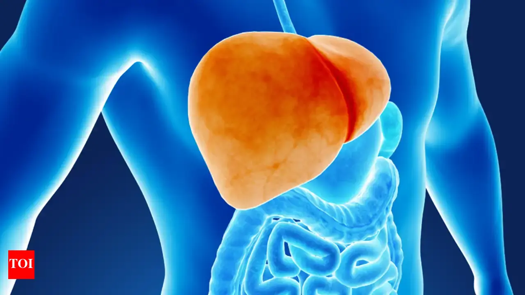
A new study from American and Korean researchers shows that DNA packaged on extracellular vesicles secreted by tumor cells can trigger an immune response that inhibits metastasis. The study, published in Nature Cancer, provides additional understanding of cancer spread and anticancer immunity, which could eventually lead to new strategies to reduce metastasis.
Extracellular vesicles are tiny capsules that cells use to exchange proteins, DNA, and other biomolecules. Tumor cells are especially active in EV secretion and prior research has shown that EVs contain DNA (EV-DNA) that represents the entire genome including cancer associated genes. The new research shows the EV-DNA secreted by tumors cells alerts anticancer immunity helping to quell liver metastases. The more EV-DNA that is present, the strong the anticancer response.
“Initially we hypothesized that more tumor EV-DNA would mean a worse prognosis, but we were surprised to find the opposite,” said study co-senior author David Lyden, MD, PhD, the Stavros S. Niarchos Professor in Pediatric Cardiology, professor of cell and developmental biology, and a member of the Sandra and Edward Meyer Cancer Center at Weill Cornell Medicine.
The mechanism of EV-DNA packaging and its role in cancer has remained elusive. The current study showed that EV-DNA is largely localized on the surface of EVs and not as “naked” strands inside the EVs.
“We had assumed that this DNA is in the form of ‘naked’ strands inside EVs, but we were surprised to find that it is mostly on the EV surface, wrapped around support proteins called histones, much as it would be in a chromosome,” said Inbal Wortzel, PhD, a postdoc working in the Lyden lab.
Working with co-senior author Yael David, PhD, an associate professor at Memorial Sloan Kettering Cancer Center, the team found that the histones had a unique set of modifications that pointed toward a specific signaling function of EV-DNA. In addition, the investigators identified a number of genes that play a role in EV-DNA packaging regulation and that when one of these genes, APAF1, was absent, the amount of secreted EV-DNA was significantly reduced.
The Lyden lab has long focused on understanding the cellular mechanisms that influence cancer metastasis. Their findings have shown that primary tumors secrete factors, including cytokines, chemokines and small extracellular vesicles (exosomes) that circulate to distant sites of future metastasis—establishing a specific microenvironment dubbed the pre-metastatic niche (PMN). The findings further demonstrate that EVs impact all aspects of the pre-metastatic niche—promoting vascular leakiness, immunosuppression, and the recruitment of pro-metastatic and pro-angiogenic bone marrow progenitor cells, which create a favorable microenvironment for the survival of tumor cells prior to their arrival at distant sites in the body.
Based on some of this earlier work, the researchers thought that EV-DNA secreted by tumors would promote metastasis, but instead found in animal models of pancreatic cancer and colorectal cancer that booting levels of tumor EV-DNA lowered metastatic risk. Further, reducing EV-DNA levels by knocking out the APAF1 gene increased the risk of metastasis.
“We also found that in colorectal cancer patients, those with low levels of EV-DNA in their tumors at the time of diagnosis were more likely to develop liver metastases later on, compared to those with high EV-DNA levels,” said co-senior author Han Sang Kim, PhD, an associate professor at the Yonsei University College of Medicine in Korea and a visiting associate professor at Weill Cornell Medicine.
Using this new information, the research intend to develop a prognostic test to measure tumor-secreted EV-DNA to assess patient risk for developing metastatic cancer. The team is also looking to develop a vaccine-like treatment that could enhance tumor EV-DNA signaling with an eye toward suppressing tumor metastasis in patients with early-stage cancer.


![Best Weight Loss Supplements [2022-23] New Reports!](https://technologytangle.com/wp-content/uploads/2022/12/p1-1170962-1670840878.png)


