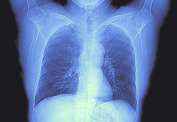
A new deep learning model has demonstrated a strong potential to accurately detect and segment lung tumors from CT scans, according to a study published in Radiology, the journal of the Radiological Society of North America (RSNA). The research suggests that the model could play a role in improving lung cancer treatment, from diagnosis to therapy planning.
“Our study represents an important step toward automating lung tumor identification and segmentation,” said lead author Mehr Kashyap, MD, a resident physician at Stanford University School of Medicine. “This approach could have wide-ranging implications, including its incorporation in automated treatment planning, tumor burden quantification, treatment response assessment and other radiomic applications.”
The study builds on previous efforts to apply artificial intelligence (AI) to lung cancer imaging, but with a significant advancement: the model was trained on a large-scale, diverse dataset, making it one of the most advanced lung tumor detection tools yet developed.
For their retrospective study, the team used a large-scale data set that of routinely collected pre-radiation treatment CT simulation scans and their associated clinical 3D segmentation. The data consisted of 1,504 CT scans with 1,828 tumor segmentations which was use to train the model, with the model then tested on 150 CT scans.
The tumor volumes predicted by the new model were compared with physician delineated volumes. Performance measurements included sensitivity, specificity, false positive rate, and Dice Similarity coefficient (DSC). DSC calculates the similarity between two sets of data by comparing the overlap between them, with a score of 0 representing no overlap and 1 perfect overlap. The model segmentations were compared to from all three physician segmentation to generate a model-physicians DSC value for each pairing.
The data showed that the model achieved a sensitivity of 92% and a specificity of 82% in detecting lung tumors, a significant improvement from earlier AI models, which often relied on manual inputs or limited datasets. For comparison, in a subset of 100 CT scans with single tumors, the model demonstrated segmentation overlap scores of 0.77, compared to a physician overlap score of 0.80. The model also completed segmentation in a fraction of the time it takes physicians.
According to the researchers, their use of a 3D U-Net architecture, helped improve the model’s performance over earlier modeling efforts, which typically used 2D models. “By capturing rich interslice information, our 3D model is theoretically capable of identifying smaller lesions that 2D models may be unable to distinguish from structures such as blood vessels and airways,” the researchers wrote.
The study’s strengths were the size and diversity of its training dataset, which were compiled from routine CT scans taken from patients undergoing radiotherapy at multiple medical centers. This allowed the model to learn from various tumor types, scanning devices, and physician segmentation techniques, making it more adaptable to different clinical settings. “The large size of our dataset and its heterogeneity are key strengths of our approach,” the researchers wrote, noting that these factors helped the model perform well on both internal and external test sets.
However, the model is not without its limitations. While it performs well on most tumors, it did not perform as well with very large tumors, often underestimating their volume. Measuring tumor volume is important information for physicians to develop a treatment plan. The researchers noted that the lack of accuracy shown by the model in assessing large tumors may have been due to larger tumors not being well-represented in the training data.
The researchers added that future studies are needed to explore how using the model could help estimate tumor burden and to evaluate treatment response over time in comparison with other models. In addition, the model’s ability to predict clinical outcomes based on its estimates of tumor burden, when combined with other prognostics models using diverse data, should also be assessed.


![Best Weight Loss Supplements [2022-23] New Reports!](https://technologytangle.com/wp-content/uploads/2022/12/p1-1170962-1670840878.png)


