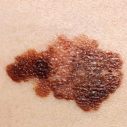
Australian researchers have shown that circulating antibodies against tumor antigens can be used as an accurate biomarker for the early detection of melanoma.
The findings were presented by Cristina Vico-Alonso, MD, lead researcher from the Victorian Melanoma Service, in Melbourne, Australia, at the European Academy of Dermatology and Venereology Congress 2024.
She told Inside Precision Medicine that a test incorporating these antibodies “would provide an earlier and more accurate diagnosis, which is essential for determining the most appropriate subsequent treatment for patients, particularly in the current scenario for melanoma, in which immunotherapy has become a game changer.”
Vico-Alonso explained that melanoma is a cancer with high mutation rate, which leads to the production of tumor antigens, known as cancer-testis antigens (CTAgs), that trigger a cellular and humoral immune response against cancer cells. The CTAg family comprises over 90 structurally and functionally diverse proteins that are aberrantly expressed in melanoma. This expression triggers the production of specific antibodies toward the relevant CTAg.
In the current study, Vico-Alonso and her team used a novel cancer-specific array developed by collaborators at the Olivia Newton John Cancer Research Institute to determine whether these circulating antibodies can reliably aid the early detection of melanoma.
They tested the array, which can detect and quantify anti-CTAg IgG antibodies against over 100 tumor antigens, on plasma samples collected from 199 patients with stage I and stage II melanoma at diagnosis and within 30 days after curative-intent surgery of the primary tumor. The results were compared with those from 38 healthy individuals without melanoma.
The analysis highlighted three tumor antigens, unique to melanoma, that show promise as diagnostic biomarkers for early-stage melanomas. The area under the curves (AUC), which indicate the level of accuracy, for each of these ranged from 0.857 to 0.981 in participants assigned to the discovery cohort, and from 0.824 to 0.985 in the internal validation cohort.
For the biomarker with an AUC value of 0.981, the corresponding sensitivity for melanoma diagnosis was 98% in the discovery cohort and 99% in the validation cohort. Specificity was and 82%, respectively.
“These results indicate that 99% of melanoma patients in the validation cohort were positive for this marker, while 82% of healthy individuals were correctly identified as negative using the recommended threshold,” explained Vico-Alonso. “While 18% of healthy individuals were incorrectly identified as positive for this marker, combining it with the other two markers into a multiparameter signature does, however, help improve accuracy.”
Based on the validation data, only one percent of melanoma patients would have a negative test for this top marker, suggesting that a negative result is highly indicative of the absence of melanoma.
Data from an external validation cohort revealed a signature of seven antigens with an AUC of 0.903, a sensitivity of 83% sensitivity, and specificity of 84% specificity.
“While this isn’t as high as in the previous cohort, it is still considered a good AUC for a diagnostic test. We are working on identifying the main confounding factors that might have contributed to these differences with the internal validation cohort,” Vico-Alonso remarked.
Prioritizing early melanoma detection is crucial because earlier diagnosis significantly improves patient outcomes, either by facilitating timely surgical intervention or by initiating systemic therapy when necessary.
Yet, melanoma diagnosis remains challenging for multiple reasons. For example, dermatologists often face difficulties in diagnosing pigmented lesions, particularly in distinguishing between benign and malignant skin lesions, while for pathologists, histopathological diagnosis can also be challenging. In certain cases, it is difficult to determine the nature of the skin lesion.
“Having additional diagnostic tools or information that offer insights into the biological significance of the skin lesion would be helpful,” said Vico-Alonso. “Additionally, we could reduce the number of unnecessary procedures like biopsies, which would be particularly beneficial for high-risk patients, such as those patients with a high nevus count.”
The researchers now plan to investigate the prognostic value of CTAgs in melanoma and also identify candidates for a pan-cancer diagnostic test, initially focusing on melanoma, lung, bowel, and pancreatic cancers.
“One significant advantage of this cancer array is its tumor-agnostic nature,” Vico-Alonso noted. “CTAgs are expressed in many solid tumors, making the diagnostic signature identified here applicable beyond melanoma. However, the specific cognate antigen combination remains unique to melanoma compared to other solid tumors.”
In addition, the team is working with professor Zongyuan Ge, PhD, from the department of data science and AI at the Monash University in Melbourne, Australia, on a multimodal AI-based approach for early detection of melanoma.
“We aim to reduce diagnostic variability and overdiagnosis by training, testing, and validating a unified multimodal AI algorithm,” said Vico-Alonso. “Our hypothesis is that integrating features from sequential macroscopic and dermoscopic images, associated histopathology, antibody profiling, and proteomic data through a multi-modal analysis in an end-to-end manner will improve AI classification accuracy and enhance melanoma diagnosis.”


